DIAGNOSIS: OSTEOSARCOMA
This is a continuation from Symptoms
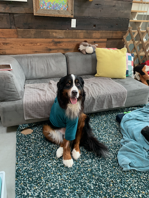
My vet could see from the x-rays that there was a tumour growing within the bone, and it was pushing the outer surface of the bone outward, hence the bowed-out lump. She asked if I would consider amputation and I said definitely. I knew Bea could handle an amputation—she is strong, fit and enthusiastic, and despite being 9 years old, she was still so agile and energetic.
People often asked me during this time how I was able to say yes to surgery so soon after the diagnosis, and it’s because as soon as I felt the lump in her leg, I knew it was cancer and started researching and preparing myself. By the time x-rays were done I’d had enough time to wrap my head around what this meant, and I knew amputation was the only viable option based on the location of the tumour. Fortunately I found the Instagram accounts of several other dogs that had recently been through the same surgery. Seeing the stages that they went through helped me to know what to expect, and I hope that my posts will help others feel a little less lost. There is a list of links to other tripod social media accounts on the Links & Resources page.
We had adopted a lab mix puppy from the SPCA about 15 years ago, and he had recently had his front leg amputated, so I had seen first hand how quickly a dog adapts to life on three legs. Arlo’s leg had been deformed from birth, so it was amputated—however in the end it turned out the leg had been deformed due to a rare type of cancer, and unfortunately because he hadn’t undergone chemotherapy, the cancer was in his lungs by age 2. Spending time with him as he learned to walk on three legs helped me make the decision about Bea having the surgery, and I knew she would be walking again soon.
SURGEON REFERRAL
We were referred to a wonderful surgeon at a nearby specialist hospital, Canada West Veterinary Specialists in Vancouver. We are fortunate enough to live only minutes from the clinic, which has made things much easier!
Dr. Hoorntje looked at the x-rays and advised that we do an immediate abdominal ultrasound. My vet had already taken chest x-rays when they did the images of the leg, otherwise a chest x-ray would have been done at this time too. This is to ensure that there was no sign of cancer elsewhere in her body, especially the lungs (which is the most common secondary site). The surgeon was also concerned that due to the unusual location on her leg bone (mid-radius) that it may be a secondary tumour, so she wanted to make sure there weren’t signs of a primary tumour elsewhere. Thankfully all looked clear, so they scheduled her for the next available date, which was September 18, 2020.
While Beatrix was sedated for the ultrasound, they also attempted a needle biopsy of the tumour. The surgeon had warned me that it can be difficult to obtain a good sample since bone tumours are extremely hard, and in the end they weren’t able to discern exactly which type of cancer we were dealing with from the biopsy. They knew it was a type of sarcoma from the sample, but were unable to say exactly what type. The final diagnosis would come after surgery, when they could take it directly from the tumour itself. They diagnosed it as osteosarcoma, which is the most common type of bone cancer in dogs.
Continued in Surgery